To study nanoscale patterns in tiny electronic or photonic components, a new method based on lensless imaging allows for near-perfect high-resolution microscopy. This is especially important at wavelengths shorter than ultraviolet, which can image with higher spatial resolution than visible light but where image-forming optics are imperfect.
The most powerful form of lensless imaging is called ptychography, which works by scanning a laser-like beam across a sample, collecting the scattered light, and then using a computer algorithm to reconstruct an image of the sample.
While ptychography can visualize many nanostructures, this special microscope has trouble analyzing samples with very regular, repeating patterns. This is because the scattered light does not change as a periodic sample is scanned, so the computer algorithm gets confused and cannot reconstruct a good image.
Taking on this challenge, recently graduated Ph.D. researchers Bin Wang and Nathan Brooks, working with JILA Fellows Margaret Murnane and Henry Kapteyn, developed a novel method that uses short-wavelength light with a special vortex or donut shape to scan these repeating surfaces, resulting in more varied diffraction patterns. This allowed the researchers to capture high-fidelity image reconstructions using this new approach, which they recently published in Optica. This result will also be highlighted in the Optica Magazine Optics and Photonics News in the annual highlights of Optics in 2023.
This new imaging method is particularly impactful for applications in nanoelectronics, photonics, and metamaterials. “The ability to structure (or change the shape of) visible laser beams into donut and other shapes has revolutionized visible super-resolution microscopy,” explained Murnane. “It is very exciting to now have a path forward for bringing these powerful capabilities to shorter wavelengths.”
Sculpting Vortex-Shaped High Harmonic Beams
To create laser-like beams at short wavelengths in a tabletop scale setup, the JILA team used a process called high harmonic generation (HHG). HHG occurs when an ultrafast laser pulse hits an atom, plucking an electron away and then driving it back to its parent atom to recombine. Upon contact, the atoms convert their electron’s kinetic energy into extreme ultraviolet (EUV) light. And if millions of atoms all emit the EUV light synchronously, the waves produce a bright laser-like EUV beam.
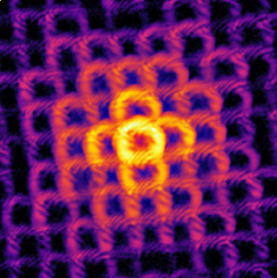
To image repeating patterns, the JILA researchers needed to find a way to change the HHG beam so that the scattered light changed as the EUV beam was scanned over the sample. The researchers coaxed the HHG beams to transform from a disk into a vortex or donut shape, which is called an orbital angular momentum (OAM) beam, to achieve this effect. This different shape would be essential to enable lensless imaging of periodic samples.
When the scientists illuminated their microscope with vortex-shaped HHG beams (see accompanying image), more intricate scatter patterns were produced, which changed as the sample was scanned. These variations encoded information about the sample’s repeating motifs and enabled the algorithm to extract an accurate image.
Beyond this exciting result, this new vortex-beam lensless imaging also produced less damage to the delicate sample than that of a scanning electron microscope. As many soft materials, plastic, and biological samples are fragile, having a precise and gentle way to image them is key.
Additionally, vortex-beam lensless imaging was better at detecting defects in the nanopattern than scanning electron microscopy, which tends to melt delicate samples.
Patterns, Patterns, Everywhere—and Now We Can See them Better
For scientists making patterned materials for next-generation nano, energy, photonic, and quantum devices, this advance enables high-resolution imaging of highly periodic structures, without destroying them. As Kapteyn elaborated: “In the future, this may also make it possible to image delicate live cells at high spatial resolution.”
Written by Kenna Hughes-Castleberry, JILA Science Communicator
 The Physics Frontiers Centers (PFC) program supports university-based centers and institutes where the collective efforts of a larger group of individuals can enable transformational advances in the most promising research areas. The program is designed to foster major breakthroughs at the intellectual frontiers of physics by providing needed resources such as combinations of talents, skills, disciplines, and/or specialized infrastructure, not usually available to individual investigators or small groups, in an environment in which the collective efforts of the larger group can be shown to be seminal to promoting significant progress in the science and the education of students. PFCs also include creative, substantive activities aimed at enhancing education, broadening participation of traditionally underrepresented groups, and outreach to the scientific community and general public.